Chronic Spine Pain?
Alleviation of chronic spine pain and headaches by reducing forward head posture and thoracic hyperkyphosis:
A CBP ® case report
Miles O Fortner 1, Paul A Oakley 2, Deed E Harrison 3
PMID: 30154612PMCID: PMC6110208 DOI: 10.1589/jpts.30.1117
August 2018
Abstract
[Purpose] This case presents the reduction of both forward head posture and thoracic hyperkyphosis in a young male with chronic back pain and headaches by a comprehensive posture rehabilitation program as a part of Chiropractic BioPhysics® methods.
[Participant and Methods] A 32 year old male presented with constant pain and headaches for seven years since he was involved in a work related injury. He had seen five different MDs, undergone multiple imaging tests, and received multiple prescriptions, thirteen steroid injections and was recommended for a spine surgery that he had denied. He was on long-term disability. Upon comprehensive posture and spine assessment, the patient had exaggerated forward head translation and thoracic hyperkyphosis. The patient was treated 36 times over 13-weeks with cervical and thoracic extension exercises, traction, and manipulation.
[Results] After treatment the patient reported dramatic improvement in symptoms as indicated on valid disability questionnaires and substantial improvements in posture.
[Conclusion] Posture-related pain and disability is not often addressed in allopathic medicine but substantial posture improvements are achievable in short time periods as this case illustrates. Poor postures in young patients should be corrected to avoid long-term consequences. Radiography as used in spinal rehabilitation is safe and reliable.
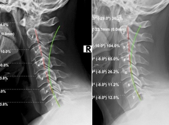
Fig. 1. Lateral cervical radiographs. Left: Initial (8/28/17); Right: Follow-up (12/6/17). Green line indicates normal ideal cervical lordosis; red line highlights patient posterior vertebral body margins (CBP Seminars, Inc.).
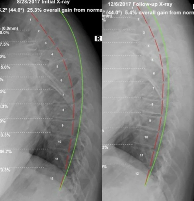
Fig. 2. Lateral thoracic radiographs. Left: Initial (8/28/17); Right: Follow-up (12/6/17). Green line indicates normal thoracic kyphosis; red line highlights patient posterior vertebral body margins (CBP Seminars, Inc.).
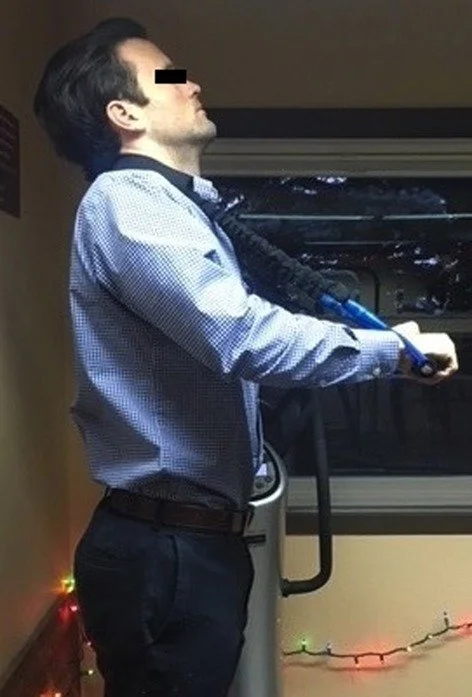
Fig. 3. Cervical mirror image hyperextension exercise.
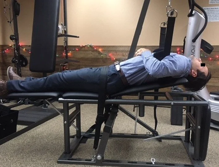
Fig. 5. Traction set-up. Patient’s thoracic spine is being forced into extension by pulling strap, and the head is allowed to be pulled by gravity backwards.
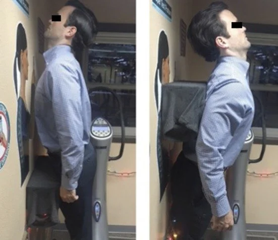
Fig. 4. Thoracic extension exercises. Left: Patient is anteriorly translating their thorax towards the wall and extending the head; Right: Patient is simultaneously retracting their head and pelvis towards the wall.
Need to contact us for an assessment to correct this type of pain or dysfunction? Call now or visit our contact page.